 Add to Chrome
 Add to Firefox
 Add to Edge
Spectrotemporal Feature Extraction in EHG Signals and Tocograms for Enhanced Preterm Birth Prediction
Sep 09, 2025Preterm birth (PTB), defined as delivery before 37 weeks of gestation, is a leading cause of neonatal mortality and long term health complications. Early detection is essential for enabling timely medical interventions. Electrohysterography (EHG) and tocography (TOCO) are promising non invasive tools for PTB prediction, but prior studies often suffer from class imbalance, improper oversampling, and reliance on features with limited physiological relevance. This work presents a machine learning pipeline incorporating robust preprocessing, physiologically grounded feature extraction, and rigorous evaluation. Features were extracted from EHG (and TOCO) signals using Mel frequency cepstral coefficients, statistical descriptors of wavelet coefficients, and peaks of the normalized power spectrum. Signal quality was enhanced via Karhunen Lo\`eve Transform (KLT) denoising through eigenvalue based subspace decomposition. Multiple classifiers, including Logistic Regression, Support Vector Machines, Random Forest, Gradient Boosting, Multilayer Perceptron, and CatBoost, were evaluated on the TPEHGT dataset. The CatBoost classifier with KLT denoising achieved the highest performance on fixed interval segments of the TPEHGT dataset, reaching 97.28% accuracy and an AUC of 0.9988. Ablation studies confirmed the critical role of both KLT denoising and physiologically informed features. Comparative analysis showed that including TOCO signals did not substantially improve prediction over EHG alone, highlighting the sufficiency of EHG for PTB detection. These results demonstrate that combining denoising with domain relevant features can yield highly accurate, robust, and clinically interpretable models, supporting the development of cost effective and accessible PTB prediction tools, particularly in low resource healthcare settings.
Assessment of Fetal and Maternal Well-Being During Pregnancy Using Passive Wearable Inertial Sensor
Nov 19, 2021


Assessing the health of both the fetus and mother is vital in preventing and identifying possible complications in pregnancy. This paper focuses on a device that can be used effectively by the mother herself with minimal supervision and provide a reasonable estimation of fetal and maternal health while being safe, comfortable, and easy to use. The device proposed uses a belt with a single accelerometer over the mother's uterus to record the required information. The device is expected to monitor both the mother and the fetus constantly over a long period and provide medical professionals with useful information, which they would otherwise overlook due to the low frequency that health monitoring is carried out at the present. The paper shows that simultaneous measurement of respiratory information of the mother and fetal movement is in fact possible even in the presence of mild interferences, which needs to be accounted for if the device is expected to be worn for extended times.
Comprehensive Study on Denoising of Medical Images Utilizing Neural Network Based Auto-Encoder
Feb 03, 2021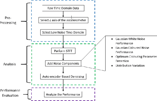
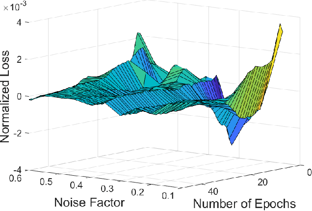
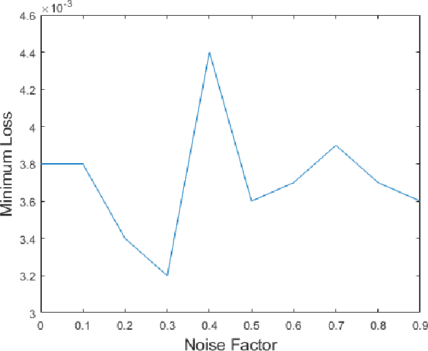
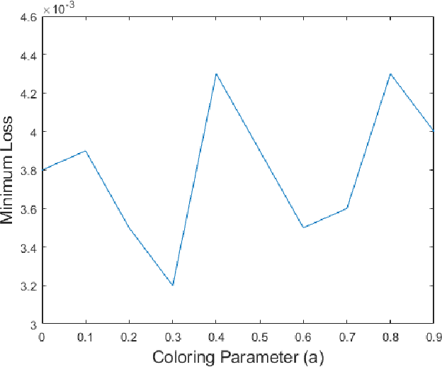
Fetal motion discernment utilizing spectral images extracted from accelerometric data incident on pregnant mothers abdomen has gained substantial attention in the state-of-the-art research. It is an essential practice to avoid adverse scenarios such as stillbirths and intrauterine growth restrictions. However, this endeavor of ensuring fetus safety has been arduous due to the existence of random noise in medical images. This novel research is an in depth approach to analyze how the interference of different noise variations affect the retrieval of information in those images. For that, an algorithm employing auto-encoder-based deep learning was modeled and the accuracy of reconstruction of the STFT images mitigating the noise has been measured examining the loss. From the results, it is manifested that even a substantial addition of the Super-Gaussian noises which have a higher correlation of the frequencies possessed by the Fetal movement images can be restored successfully with the slightest error.
Novel Non-Invasive In-house Fabricated Wearable System with a Hybrid Algorithm for Fetal Movement Recognition
Jan 29, 2021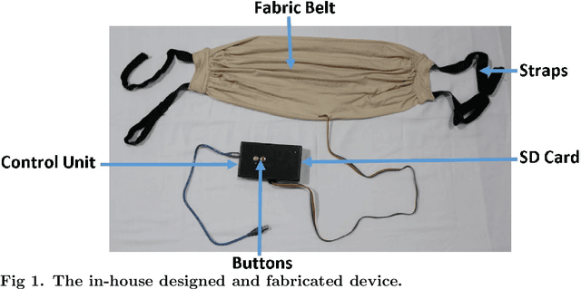
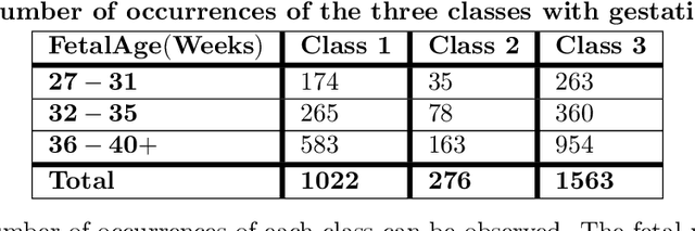
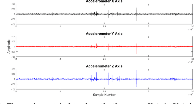
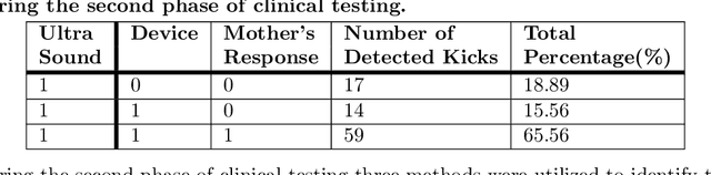
Fetal movement count monitoring is one of the most commonly used methods of assessing fetal well-being. While few methods are available to monitor fetal movements, they consist of several adverse qualities such as unreliability as well as the inability to be conducted in a non-clinical setting. Therefore, this research was conducted to design a complete system that will enable pregnant mothers to monitor fetal movement at home. This system consists of a non-invasive, non-transmitting sensor unit that can be fabricated at a low cost. An accelerometer was utilized as the primary sensor and a micro-controller based circuit was implemented. Clinical testing was conducted utilizing this sensor unit. Two phases of clinical testing procedures were done and readings from more than 120 pregnant mothers were taking. Validation was done by conducting an abdominal ultrasound scan which was utilized as the ground truth during the second phase of the clinical testing procedure. A clinical survey was also conducted in parallel with clinical testings in order to improve the sensor unit as well as to improve the final system. Four different signal processing algorithms were implemented on the data set and the performance of each was compared with each other. Consequently, the most feasible as well as the best performing algorithm was determined and it was utilized in the final system. Furthermore, a mobile application was also developed to be used with the sensor unit by pregnant mothers. Finally, a complete end to end method to monitor fetal movement in a non-clinical setting was presented by the proposed system.