Aravindan Chandrabose
Few-Shot Classification of Skin Lesions from Dermoscopic Images by Meta-Learning Representative Embeddings
Oct 30, 2022Abstract:Annotated images and ground truth for the diagnosis of rare and novel diseases are scarce. This is expected to prevail, considering the small number of affected patient population and limited clinical expertise to annotate images. Further, the frequently occurring long-tailed class distributions in skin lesion and other disease classification datasets cause conventional training approaches to lead to poor generalization due to biased class priors. Few-shot learning, and meta-learning in general, aim to overcome these issues by aiming to perform well in low data regimes. This paper focuses on improving meta-learning for the classification of dermoscopic images. Specifically, we propose a baseline supervised method on the meta-training set that allows a network to learn highly representative and generalizable feature embeddings for images, that are readily transferable to new few-shot learning tasks. We follow some of the previous work in literature that posit that a representative feature embedding can be more effective than complex meta-learning algorithms. We empirically prove the efficacy of the proposed meta-training method on dermoscopic images for learning embeddings, and show that even simple linear classifiers trained atop these representations suffice to outperform some of the usual meta-learning methods.
Deep Learning for Skin Lesion Classification
Mar 13, 2017


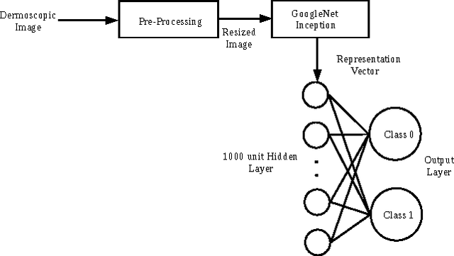
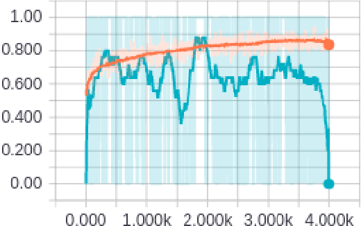
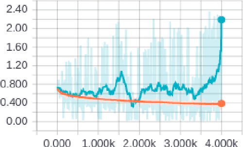
Abstract:Melanoma, a malignant form of skin cancer is very threatening to life. Diagnosis of melanoma at an earlier stage is highly needed as it has a very high cure rate. Benign and malignant forms of skin cancer can be detected by analyzing the lesions present on the surface of the skin using dermoscopic images. In this work, an automated skin lesion detection system has been developed which learns the representation of the image using Google's pretrained CNN model known as Inception-v3 \cite{cnn}. After obtaining the representation vector for our input dermoscopic images we have trained two layer feed forward neural network to classify the images as malignant or benign. The system also classifies the images based on the cause of the cancer either due to melanocytic or non-melanocytic cells using a different neural network. These classification tasks are part of the challenge organized by International Skin Imaging Collaboration (ISIC) 2017. Our system learns to classify the images based on the model built using the training images given in the challenge and the experimental results were evaluated using validation and test sets. Our system has achieved an overall accuracy of 65.8\% for the validation set.
Automatic Skin Lesion Segmentation using Semi-supervised Learning Technique
Mar 13, 2017
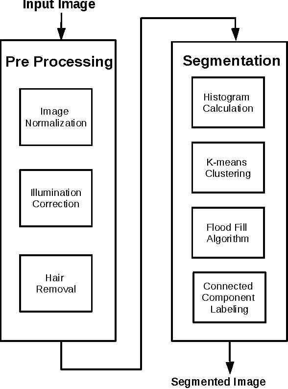
Abstract:Skin cancer is the most common of all cancers and each year million cases of skin cancer are treated. Treating and curing skin cancer is easy, if it is diagnosed and treated at an early stage. In this work we propose an automatic technique for skin lesion segmentation in dermoscopic images which helps in classifying the skin cancer types. The proposed method comprises of two major phases (1) preprocessing and (2) segmentation using semi-supervised learning algorithm. In the preprocessing phase noise are removed using filtering technique and in the segmentation phase skin lesions are segmented based on clustering technique. K-means clustering algorithm is used to cluster the preprocessed images and skin lesions are filtered from these clusters based on the color feature. Color of the skin lesions are learned from the training images using histograms calculations in RGB color space. The training images were downloaded from the ISIC 2017 challenge website and the experimental results were evaluated using validation and test sets.
 Add to Chrome
 Add to Firefox
 Add to Edge