Alnur Alimanov
Denoising Diffusion Probabilistic Model for Retinal Image Generation and Segmentation
Aug 16, 2023Abstract:Experts use retinal images and vessel trees to detect and diagnose various eye, blood circulation, and brain-related diseases. However, manual segmentation of retinal images is a time-consuming process that requires high expertise and is difficult due to privacy issues. Many methods have been proposed to segment images, but the need for large retinal image datasets limits the performance of these methods. Several methods synthesize deep learning models based on Generative Adversarial Networks (GAN) to generate limited sample varieties. This paper proposes a novel Denoising Diffusion Probabilistic Model (DDPM) that outperformed GANs in image synthesis. We developed a Retinal Trees (ReTree) dataset consisting of retinal images, corresponding vessel trees, and a segmentation network based on DDPM trained with images from the ReTree dataset. In the first stage, we develop a two-stage DDPM that generates vessel trees from random numbers belonging to a standard normal distribution. Later, the model is guided to generate fundus images from given vessel trees and random distribution. The proposed dataset has been evaluated quantitatively and qualitatively. Quantitative evaluation metrics include Frechet Inception Distance (FID) score, Jaccard similarity coefficient, Cohen's kappa, Matthew's Correlation Coefficient (MCC), precision, recall, F1-score, and accuracy. We trained the vessel segmentation model with synthetic data to validate our dataset's efficiency and tested it on authentic data. Our developed dataset and source code is available at https://github.com/AAleka/retree.
Retinal Image Restoration using Transformer and Cycle-Consistent Generative Adversarial Network
Mar 03, 2023Abstract:Medical imaging plays a significant role in detecting and treating various diseases. However, these images often happen to be of too poor quality, leading to decreased efficiency, extra expenses, and even incorrect diagnoses. Therefore, we propose a retinal image enhancement method using a vision transformer and convolutional neural network. It builds a cycle-consistent generative adversarial network that relies on unpaired datasets. It consists of two generators that translate images from one domain to another (e.g., low- to high-quality and vice versa), playing an adversarial game with two discriminators. Generators produce indistinguishable images for discriminators that predict the original images from generated ones. Generators are a combination of vision transformer (ViT) encoder and convolutional neural network (CNN) decoder. Discriminators include traditional CNN encoders. The resulting improved images have been tested quantitatively using such evaluation metrics as peak signal-to-noise ratio (PSNR), structural similarity index measure (SSIM), and qualitatively, i.e., vessel segmentation. The proposed method successfully reduces the adverse effects of blurring, noise, illumination disturbances, and color distortions while significantly preserving structural and color information. Experimental results show the superiority of the proposed method. Our testing PSNR is 31.138 dB for the first and 27.798 dB for the second dataset. Testing SSIM is 0.919 and 0.904, respectively.
Retinal Image Restoration and Vessel Segmentation using Modified Cycle-CBAM and CBAM-UNet
Sep 09, 2022


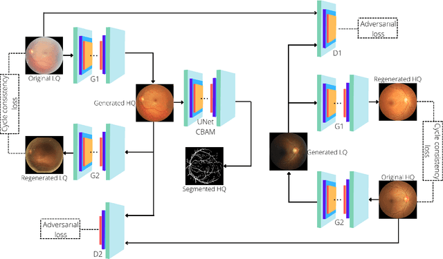
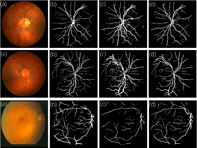
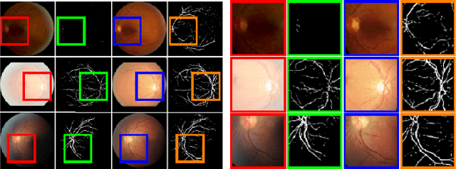
Abstract:Clinical screening with low-quality fundus images is challenging and significantly leads to misdiagnosis. This paper addresses the issue of improving the retinal image quality and vessel segmentation through retinal image restoration. More specifically, a cycle-consistent generative adversarial network (CycleGAN) with a convolution block attention module (CBAM) is used for retinal image restoration. A modified UNet is used for retinal vessel segmentation for the restored retinal images (CBAM-UNet). The proposed model consists of two generators and two discriminators. Generators translate images from one domain to another, i.e., from low to high quality and vice versa. Discriminators classify generated and original images. The retinal vessel segmentation model uses downsampling, bottlenecking, and upsampling layers to generate segmented images. The CBAM has been used to enhance the feature extraction of these models. The proposed method does not require paired image datasets, which are challenging to produce. Instead, it uses unpaired data that consists of low- and high-quality fundus images retrieved from publicly available datasets. The restoration performance of the proposed method was evaluated using full-reference evaluation metrics, e.g., peak signal-to-noise ratio (PSNR) and structural similarity index measure (SSIM). The retinal vessel segmentation performance was compared with the ground-truth fundus images. The proposed method can significantly reduce the degradation effects caused by out-of-focus blurring, color distortion, low, high, and uneven illumination. Experimental results show the effectiveness of the proposed method for retinal image restoration and vessel segmentation.
 Add to Chrome
 Add to Firefox
 Add to Edge