Towards Non-contact Muscle Activity Estimation using FMCW Radar
Paper and Code
Dec 21, 2023


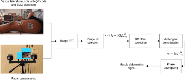
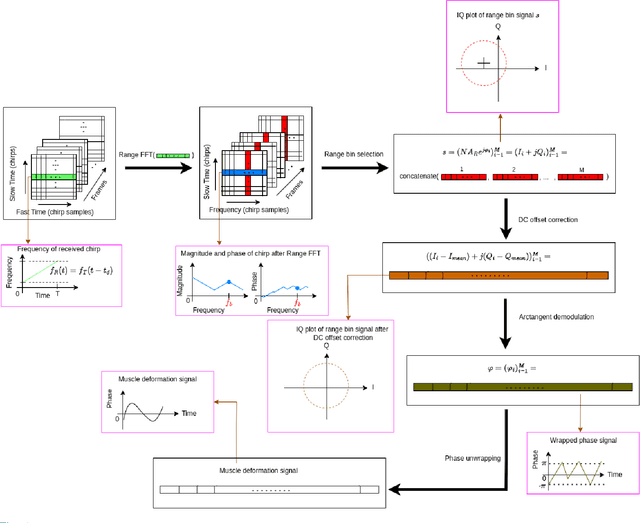
Surface electromyography (sEMG) is a widely used muscle activity monitoring technique. sEMG measures muscle activity through monopolar and bipolar, multi-electrode electrodes. The surface electrodes are placed on the surface of the skin above the target muscle and the received signal can be used to infer the state of the muscle - active, inactive or fatigued - which serves as vital information during neurological and orthopaedic rehabilitation. Additionally, the sEMG signal can also be used for the control of prostheses. sEMG requires contact with the participant's skin and is thus a potentially uncomfortable method for the measurement of muscle activity. Moreover, the setup procedure has been termed time-consuming by sEMG experts and is listed as one of the main barriers to the clinical employment of the technique. Previous studies have shown that architectural changes, particularly muscle deformation, can provide information about the activity of the muscle, providing an alternative to sEMG. In all these studies, the muscle deformation signal is acquired using ultrasound imaging, an approach known as sonomyography (SMG). Despite its advantages, such as improved spatial resolution, SMG is still a contact based approach. In this paper, we propose a non-contact muscle activity monitoring approach that measures the muscle deformation signal using a Frequency Modulated Continuous Wave (FMCW) mmWave radar which we call radiomyography (RMG). In future, this system will enable muscle activation to be measured in an unconstrained and less cumbersome manner for both the person conducting the test and the individual being tested.
 Add to Chrome
 Add to Firefox
 Add to Edge