Simultaneous column-based deep learning progression analysis of atrophy associated with AMD in longitudinal OCT studies
Paper and Code
Jul 31, 2023


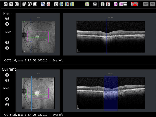
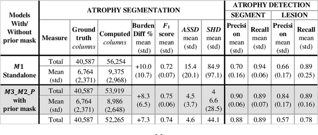
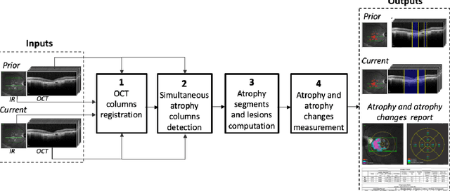
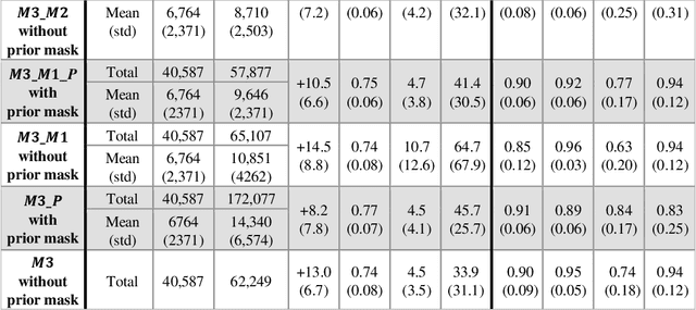
Purpose: Disease progression of retinal atrophy associated with AMD requires the accurate quantification of the retinal atrophy changes on longitudinal OCT studies. It is based on finding, comparing, and delineating subtle atrophy changes on consecutive pairs (prior and current) of unregistered OCT scans. Methods: We present a fully automatic end-to-end pipeline for the simultaneous detection and quantification of time-related atrophy changes associated with dry AMD in pairs of OCT scans of a patient. It uses a novel simultaneous multi-channel column-based deep learning model trained on registered pairs of OCT scans that concurrently detects and segments retinal atrophy segments in consecutive OCT scans by classifying light scattering patterns in matched pairs of vertical pixel-wide columns (A-scans) in registered prior and current OCT slices (B-scans). Results: Experimental results on 4,040 OCT slices with 5.2M columns from 40 scans pairs of 18 patients (66% training/validation, 33% testing) with 24.13+-14.0 months apart in which Complete RPE and Outer Retinal Atrophy (cRORA) was identified in 1,998 OCT slices (735 atrophy lesions from 3,732 segments, 0.45M columns) yield a mean atrophy segments detection precision, recall of 0.90+-0.09, 0.95+-0.06 and 0.74+-0.18, 0.94+-0.12 for atrophy lesions with AUC=0.897, all above observer variability. Simultaneous classification outperforms standalone classification precision and recall by 30+-62% and 27+-0% for atrophy segments and lesions. Conclusions: simultaneous column-based detection and quantification of retinal atrophy changes associated with AMD is accurate and outperforms standalone classification methods. Translational relevance: an automatic and efficient way to detect and quantify retinal atrophy changes associated with AMD.
 Add to Chrome
 Add to Firefox
 Add to Edge