Separating Overlapping Tissue Layers from Microscopy Images
Paper and Code
May 22, 2019


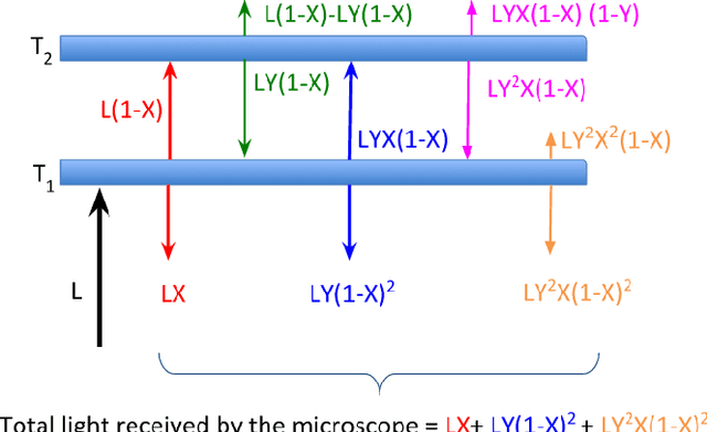
Manual preparation of tissue slices for microscopy imaging can introduce tissue tears and overlaps. Typically, further digital processing algorithms such as registration and 3D reconstruction from tissue image stacks cannot handle images with tissue tear/overlap artifacts, and so such images are usually discarded. In this paper, we propose an imaging model and an algorithm to digitally separate overlapping tissue data of mouse brain images into two layers. We show the correctness of our model and the algorithm by comparing our results with the ground truth.
 Add to Chrome
 Add to Firefox
 Add to Edge