Non-invasive Localization of the Ventricular Excitation Origin Without Patient-specific Geometries Using Deep Learning
Paper and Code
Sep 16, 2022


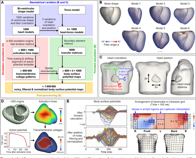
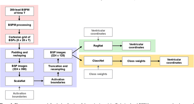
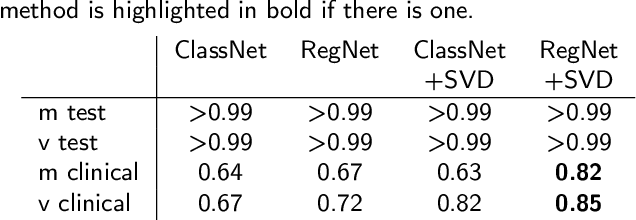
Ventricular tachycardia (VT) can be one cause of sudden cardiac death affecting 4.25 million persons per year worldwide. A curative treatment is catheter ablation in order to inactivate the abnormally triggering regions. To facilitate and expedite the localization during the ablation procedure, we present two novel localization techniques based on convolutional neural networks (CNNs). In contrast to existing methods, e.g. using ECG imaging, our approaches were designed to be independent of the patient-specific geometries and directly applicable to surface ECG signals, while also delivering a binary transmural position. One method outputs ranked alternative solutions. Results can be visualized either on a generic or patient geometry. The CNNs were trained on a data set containing only simulated data and evaluated both on simulated and clinical test data. On simulated data, the median test error was below 3mm. The median localization error on the clinical data was as low as 32mm. The transmural position was correctly detected in up to 82% of all clinical cases. Using the ranked alternative solutions, the top-3 median error dropped to 20mm on clinical data. These results demonstrate a proof of principle to utilize CNNs to localize the activation source without the intrinsic need of patient-specific geometrical information. Furthermore, delivering multiple solutions can help the physician to find the real activation source amongst more than one possible locations. With further optimization, these methods have a high potential to speed up clinical interventions. Consequently they could decrease procedural risk and improve VT patients' outcomes.
 Add to Chrome
 Add to Firefox
 Add to Edge