Classification of anatomic structures in head and neck by CT-based radiomics
Paper and Code
May 17, 2022


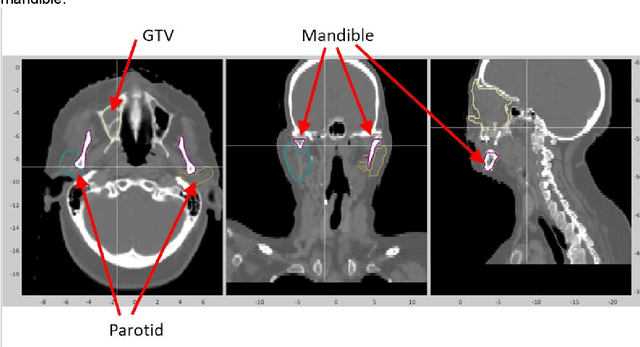
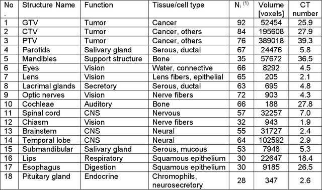
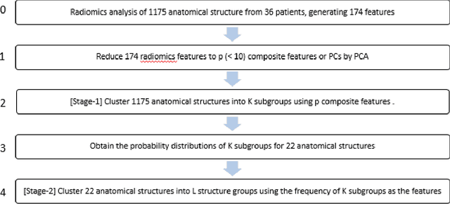
Background and Purpose: Radiomics features are used to identify disease types and predict therapy outcomes. However, how the radiomics features are different among different anatomical structures has never been investigated. Hence, we analyzed the radiomics features of 22 anatomical structures in the head and neck area in CT images. Furthermore, we studied whether CT radiomics can classify anatomical structures of the head and neck using unsupervised machine-learning techniques. Materials and methods: We obtained IMRT/VMAT treatment planning data from 36 patients treated for head and neck cancers in a single institution. There were 1357 contours of more than 22 anatomical structures drawn on planning CTs. We calculated 174 radiomics features using the SIBEX program. First, we tested whether the radiomics features of anatomical structures were unique enough to classify all contours into 22 groups. We then developed a two-stage clustering technique to classify 22 anatomic structures into sub-groups with similar physiological or biological characteristics. Results: The heatmap of 174 radiomics features of 22 anatomical structures showed a distinct difference among tumors and other healthy structures. Radiomics features have allowed us to identify the eyes, lens, submandibular, pituitary glands, and thyroids with over 90% accuracy. The two-stage clustering of 22 structures resulted in six subgroups, which shared common characteristics such as fatty and bony tissues. Conclusions: We have shown that anatomical structures in head and neck tumors have distinguishable radiomics features. We could observe similarities of features among subgroups of the structures. The results suggest that CT radiomics can help distinguish the biological characteristics of head and neck lesions.
 Add to Chrome
 Add to Firefox
 Add to Edge