 Add to Chrome
 Add to Firefox
 Add to Edge
An AI-IoT Based Smart Wheelchair with Gesture-Controlled Mobility, Deep Learning-Based Obstacle Detection, Multi-Sensor Health Monitoring, and Emergency Alert System
Jan 17, 2026The growing number of differently-abled and elderly individuals demands affordable, intelligent wheelchairs that combine safe navigation with health monitoring. Traditional wheelchairs lack dynamic features, and many smart alternatives remain costly, single-modality, and limited in health integration. Motivated by the pressing demand for advanced, personalized, and affordable assistive technologies, we propose a comprehensive AI-IoT based smart wheelchair system that incorporates glove-based gesture control for hands-free navigation, real-time object detection using YOLOv8 with auditory feedback for obstacle avoidance, and ultrasonic for immediate collision avoidance. Vital signs (heart rate, SpO$_2$, ECG, temperature) are continuously monitored, uploaded to ThingSpeak, and trigger email alerts for critical conditions. Built on a modular and low-cost architecture, the gesture control achieved a 95.5\% success rate, ultrasonic obstacle detection reached 94\% accuracy, and YOLOv8-based object detection delivered 91.5\% Precision, 90.2\% Recall, and a 90.8\% F1-score. This integrated, multi-modal approach offers a practical, scalable, and affordable solution, significantly enhancing user autonomy, safety, and independence by bridging the gap between innovative research and real-world deployment.
Osteosarcoma Tumor Detection using Transfer Learning Models
May 16, 2023


The field of clinical image analysis has been applying transfer learning models increasingly due to their less computational complexity, better accuracy etc. These are pre-trained models that don't require to be trained from scratch which eliminates the necessity of large datasets. Transfer learning models are mostly used for the analysis of brain, breast, or lung images but other sectors such as bone marrow cell detection or bone cancer detection can also benefit from using transfer learning models, especially considering the lack of available large datasets for these tasks. This paper studies the performance of several transfer learning models for osteosarcoma tumour detection. Osteosarcoma is a type of bone cancer mostly found in the cells of the long bones of the body. The dataset consists of H&E stained images divided into 4 categories- Viable Tumor, Non-viable Tumor, Non-Tumor and Viable Non-viable. Both datasets were randomly divided into train and test sets following an 80-20 ratio. 80% was used for training and 20\% for test. 4 models are considered for comparison- EfficientNetB7, InceptionResNetV2, NasNetLarge and ResNet50. All these models are pre-trained on ImageNet. According to the result, InceptionResNetV2 achieved the highest accuracy (93.29%), followed by NasNetLarge (90.91%), ResNet50 (89.83%) and EfficientNetB7 (62.77%). It also had the highest precision (0.8658) and recall (0.8658) values among the 4 models.
Bone Marrow Cytomorphology Cell Detection using InceptionResNetV2
May 09, 2023
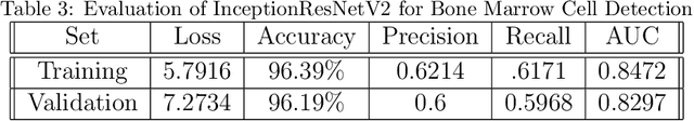
Critical clinical decision points in haematology are influenced by the requirement of bone marrow cytology for a haematological diagnosis. Bone marrow cytology, however, is restricted to reference facilities with expertise, and linked to inter-observer variability which requires a long time to process that could result in a delayed or inaccurate diagnosis, leaving an unmet need for cutting-edge supporting technologies. This paper presents a novel transfer learning model for Bone Marrow Cell Detection to provide a solution to all the difficulties faced for the task along with considerable accuracy. The proposed model achieved 96.19\% accuracy which can be used in the future for analysis of other medical images in this domain.