Irteza Enan Kabir
Angular Integral Autocorrelation for Speed Estimation in Shear Wave Elastography
Feb 02, 2024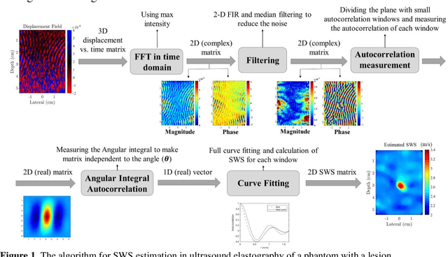

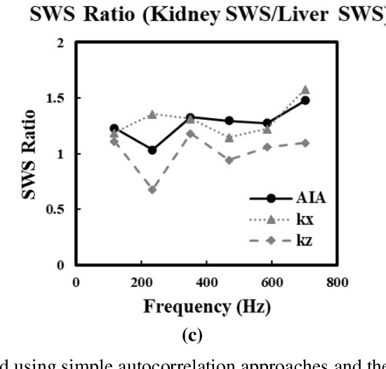

Abstract:The utilization of a reverberant shear wave field in shear wave elastography has emerged as a promising technique for achieving robust shear wave speed (SWS) estimation. However, accurately measuring SWS within such a complex wave field remains challenging. This study introduces an advanced autocorrelation estimator based on angular integration known as the angular integral autocorrelation (AIA) approach to address this issue. The AIA approach incorporates all the au-tocorrelation data from various angles during measurements, resulting in enhanced robustness to both noise and imperfect distributions in SWS estimation. The effectiveness of the AIA estimator for SWS estimation is first validated using a k-Wave simulation of a stiff branching tube in a uniform background. Furthermore, the AIA estimator is applied to ultrasound elastography experiments, MRI experiments, and OCT studies across a range of different excitation frequencies on tissues and phantoms, including in vivo scans. The results verify the capacity of the AIA approach to enhance the accuracy of SWS estimation and SNR, even within an imperfect reverberant shear wave field. Compared to simple autocorrelation approaches, the AIA approach can also successfully visualize and define lesions while significantly improving the estimated SWS and SNR in homogeneous background materials, thus providing improved elastic contrast between structures within the scans. These findings demonstrate the robustness and effectiveness of the AIA approach across a wide range of applications, including ultrasound elastography, MRE, and OCE, for accurately identifying the elastic properties of biological tissues in diverse excitation scenarios.
Imrpoving Strain Estimation in Breast Ultrasound Images Using Novel 1.5D Approach (Simulation and In-vivo results
Oct 13, 2022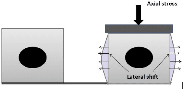
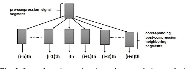
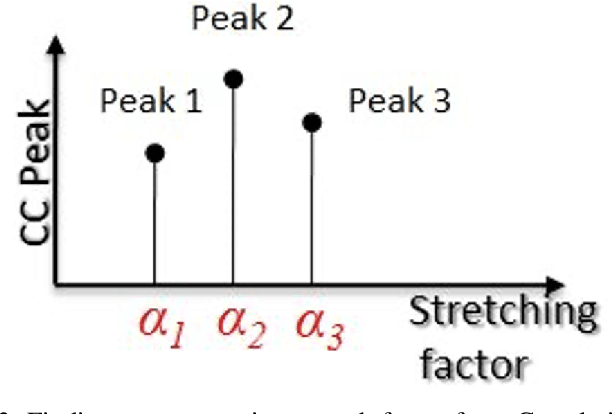
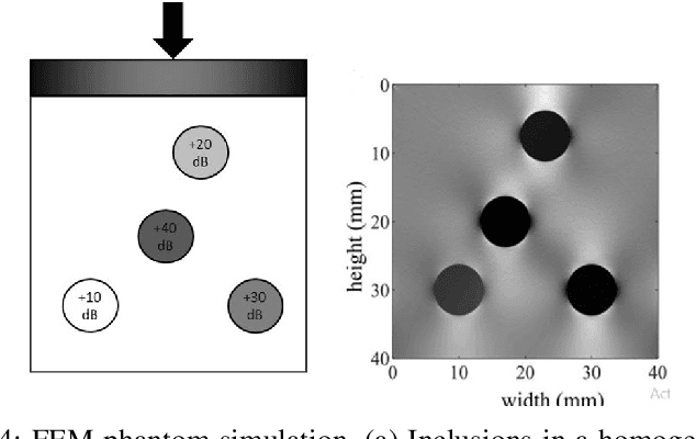
Abstract:Ultrasound elastography is the method to image the elasticity of compliant tissues due to a mechanical compression applied to it. In elastography, the local strain of explored tissue is estimated by analyzing the echo signals. This is accomplished by a sonographer who uses ultrasound transducer to apply pressure on the tissue area causing displacement of the tissue. A set of data is obtained before and after the compression. This stress along the axial direction not only causes deformation in that direction but it can also cause lateral deformation in the tissue. This relative deformation is later calculated using tracking algorithms. Finally, estimating the elasticity, images are formed. In this paper, we introduce a novel strain estimator. The proposed 1.5D strain estimator uses 1D windows for swift computations and searches the lateral direction for taking the non-axial movement into account. We have investigated the performance of our estimator using simulated and in-vivo data of breast tissue. The performance of our method is verified by comparing with different methods up to 16\% applied strain. The proposed estimator has exhibited a superior performance compared to the conventional methods. The image quality significantly improves, especially in the presence of higher applied strain, when the non-axial motion is significant.
Gastrointestinal Disorder Detection with a Transformer Based Approach
Oct 06, 2022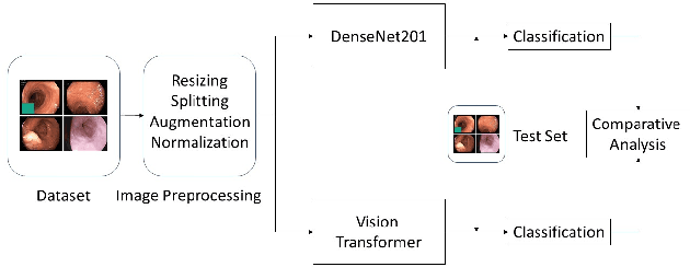
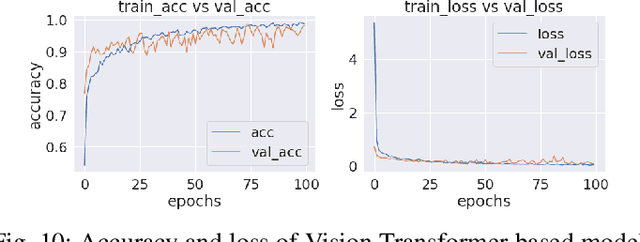
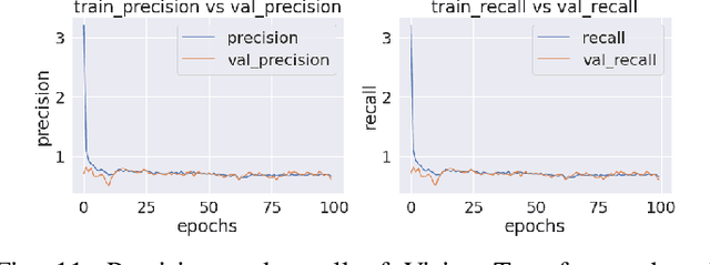
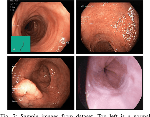
Abstract:Accurate disease categorization using endoscopic images is a significant problem in Gastroenterology. This paper describes a technique for assisting medical diagnosis procedures and identifying gastrointestinal tract disorders based on the categorization of characteristics taken from endoscopic pictures using a vision transformer and transfer learning model. Vision transformer has shown very promising results on difficult image classification tasks. In this paper, we have suggested a vision transformer based approach to detect gastrointestianl diseases from wireless capsule endoscopy (WCE) curated images of colon with an accuracy of 95.63\%. We have compared this transformer based approach with pretrained convolutional neural network (CNN) model DenseNet201 and demonstrated that vision transformer surpassed DenseNet201 in various quantitative performance evaluation metrics.
PCONet: A Convolutional Neural Network Architecture to Detect Polycystic Ovary Syndrome (PCOS) from Ovarian Ultrasound Images
Oct 02, 2022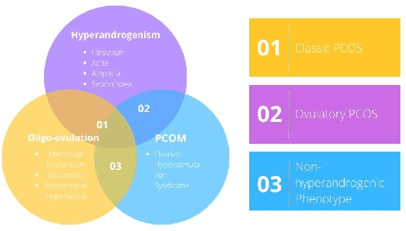


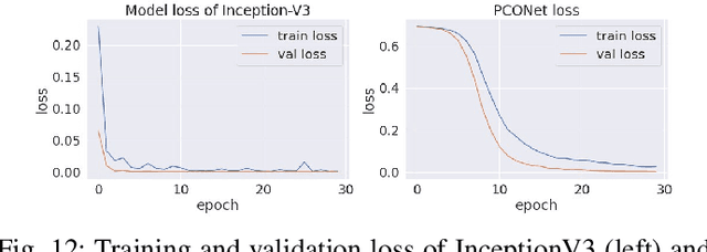
Abstract:Polycystic Ovary Syndrome (PCOS) is an endrocrinological dysfunction prevalent among women of reproductive age. PCOS is a combination of syndromes caused by an excess of androgens - a group of sex hormones - in women. Syndromes including acne, alopecia, hirsutism, hyperandrogenaemia, oligo-ovulation, etc. are caused by PCOS. It is also a major cause of female infertility. An estimated 15% of reproductive-aged women are affected by PCOS globally. The necessity of detecting PCOS early due to the severity of its deleterious effects cannot be overstated. In this paper, we have developed PCONet - a Convolutional Neural Network (CNN) - to detect polycistic ovary from ovarian ultrasound images. We have also fine tuned InceptionV3 - a pretrained convolutional neural network of 45 layers - by utilizing the transfer learning method to classify polcystic ovarian ultrasound images. We have compared these two models on various quantitative performance evaluation parameters and demonstrated that PCONet is the superior one among these two with an accuracy of 98.12%, whereas the fine tuned InceptionV3 showcased an accuracy of 96.56% on test images.
 Add to Chrome
 Add to Firefox
 Add to Edge