Andrea Farina
Initial non-invasive in vivo sensing of the lung using time domain diffuse optics
May 17, 2022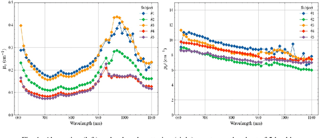
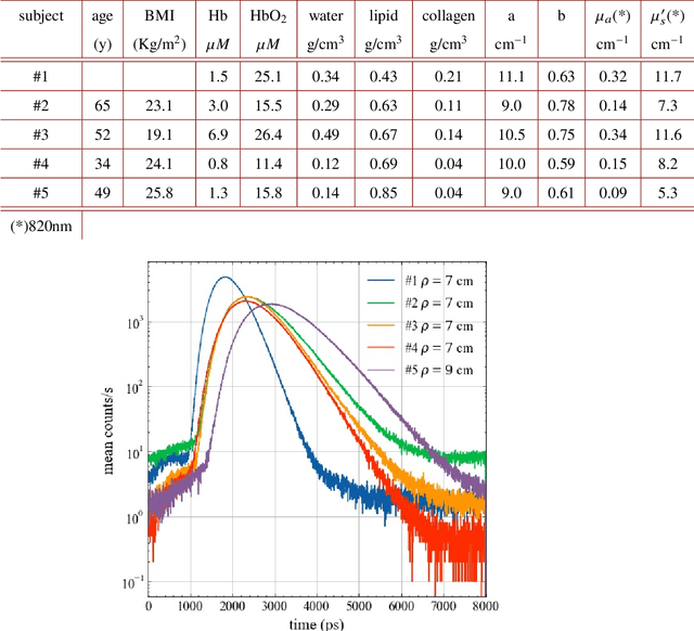
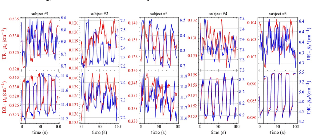
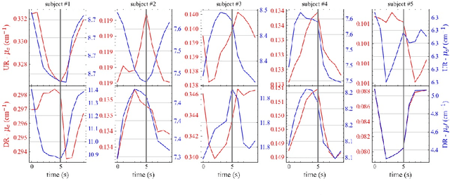
Abstract:Non-invasive in vivo sensing of the lung with light would help diagnose and monitor pulmonary disorders (caused by e.g. COVID-19, emphysema, immature lung tissue in infants). We investigated the possibility to probe the lung with time domain diffuse optics, taking advantage of the increased depth (few cm) reached by photons detected after a long (few ns) propagation time. An initial study on 5 healthy volunteers included time-resolved broadband diffuse optical spectroscopy measurements at 3 cm source-detector distance over the 600-1100 nm range, and long-distance (6-9 cm) measurements at 820 nm performed during a breathing protocol. The interpretation of the in vivo data with a simplified homogeneous model yielded a maximum probing depth of 2.6-3.9 cm, suitable to reach the lung. Also, signal changes related to the inspiration act were observed, especially at high photon propagation times. Yet, intra- and inter-subject variability and inconsistencies, possibly alluring to competing scattering and absorption effects, prevented a simple interpretation. Aspects to be further investigated to gain a deeper insight are discussed.
Giga-voxel multidimensional fluorescence imaging combining single-pixel detection and data fusion
Jul 26, 2021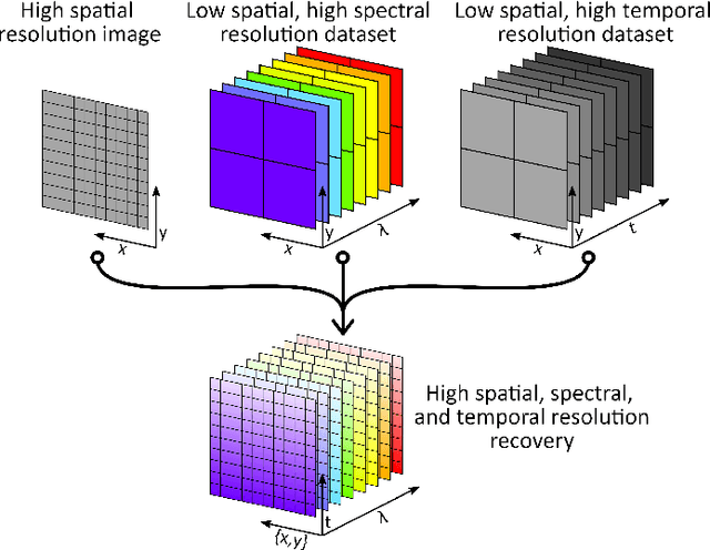
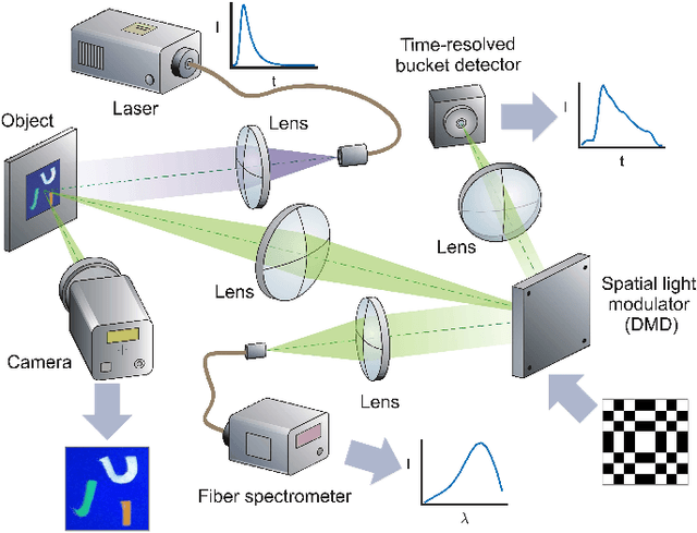
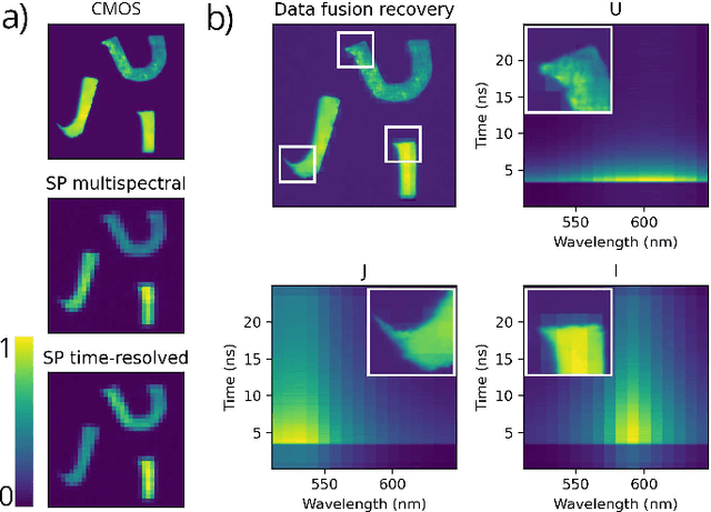
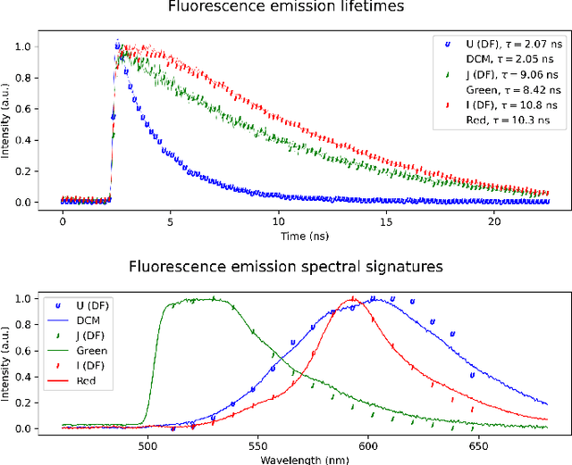
Abstract:Time-resolved fluorescence imaging is a key tool in biomedical applications, as it allows to non-invasively obtain functional and structural information. However, the big amount of collected data introduces challenges in both acquisition speed and processing needs. Here, we introduce a novel technique that allows to reconstruct a Giga-voxel 4D hypercube in a fast manner while only measuring 0.03 % of the information. The system combines two single-pixel cameras and a conventional 2D array detector working in parallel. Data fusion techniques are introduced to combine the individual 2D and 3D projections acquired by each sensor in the final high-resolution 4D hypercube, which can be used to identify different fluorophore species by their spectral and temporal signatures.
 Add to Chrome
 Add to Firefox
 Add to Edge