Aaron Chung-Jukko
Virtual Extended-Range Tomography (VERT): Contact-free realistic ultrasonic bone imaging
May 05, 2024


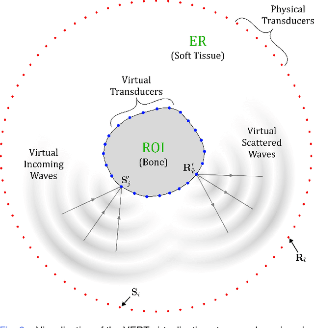
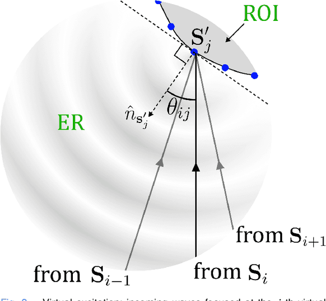
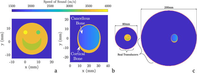
Abstract:Ultrasound tomography generally struggles to reconstruct high-contrast and/or extended-range problems. A prime example is site-specific in-vivo bone imaging, crucial for accurately assessing the risk of life-threatening fractures, which are preventable given accurate diagnosis and treatment. In this type of problem, two main obstacles arise: (a) an external region prohibits access to the region of interest (ROI), and (b) high contrast exists between the two regions. These challenges impede existing algorithms -- including bent-ray tomography (BRT), known for its robustness, speed, and reasonable short-range resolution. We propose Virtual Extended-Range Tomography (VERT), which tackles these challenges through (a) placement of virtual transducers directly on the ROI, facilitating (b) rapid initialisation before BRT inversion. In-silico validation against BRT with and without a-priori information shows superior resolution and robustness -- while maintaining or even improving speed. These improvements are drastic where the external region is much larger than the ROI. Additional validation against the practically impossible -- BRT directly on the ROI -- demonstrates that VERT is approaching the resolution limit. The capability to solve high-contrast extended-range tomography problems without prior knowledge about the ROI's interior has many implications. VERT has the potential to unlock site-specific in-vivo bone imaging for assessing fracture risk, potentially saving millions of lives globally. In other applications, VERT may replace classical BRT to yield improvements in resolution, robustness and speed -- especially where the ROI does not cover the entire imaging array. For even higher resolution, VERT offers a reliable starting background to complement algorithms with less robustness and high computational costs.
 Add to Chrome
 Add to Firefox
 Add to Edge