Improved Diagnosis of Tibiofemoral Cartilage Defects on MRI Images Using Deep Learning
Paper and Code
Nov 30, 2020


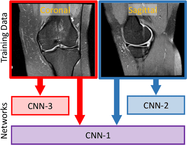
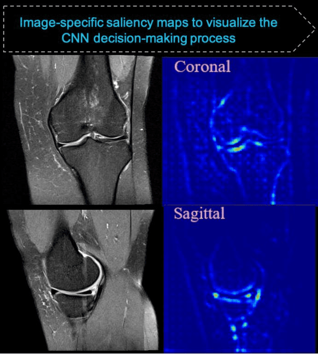
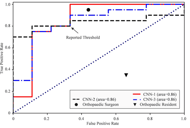
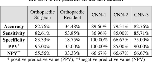
Background: MRI is the modality of choice for cartilage imaging; however, its diagnostic performance is variable and significantly lower than the gold standard diagnostic knee arthroscopy. In recent years, deep learning has been used to automatically interpret medical images to improve diagnostic accuracy and speed. Purpose: The primary purpose of this study was to evaluate whether deep learning applied to the interpretation of knee MRI images can be utilized to identify cartilage defects accurately. Methods: We analyzed data from patients who underwent knee MRI evaluation and consequently had arthroscopic knee surgery (207 with cartilage defect, 90 without cartilage defect). Patients' arthroscopic findings were compared to preoperative MRI images to verify the presence or absence of isolated tibiofemoral cartilage defects. We developed three convolutional neural networks (CNNs) to analyze the MRI images and implemented image-specific saliency maps to visualize the CNNs' decision-making process. To compare the CNNs' performance against human interpretation, the same test dataset images were provided to an experienced orthopaedic surgeon and an orthopaedic resident. Results: Saliency maps demonstrated that the CNNs learned to focus on the clinically relevant areas of the tibiofemoral articular cartilage on MRI images during the decision-making processes. One CNN achieved higher performance than the orthopaedic surgeon, with two more accurate diagnoses made by the CNN. All the CNNs outperformed the orthopaedic resident. Conclusion: CNN can be used to enhance the diagnostic performance of MRI in identifying isolated tibiofemoral cartilage defects and may replace diagnostic knee arthroscopy in certain cases in the future.
 Add to Chrome
 Add to Firefox
 Add to Edge